HUMERUS FRACTURES

By: Assoc. Dr. ancestor
HUMERUS FRACTURES
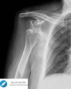
Upper end fractures of the humerus usually occur due to indirect trauma. It constitutes %4-5 of all fractures and mostly occurs as a result of falling on the hand while the arm is abducted. It accounts for % 45% of humerus fractures and is common in women over 40 years of age. There is minimal displacement in approximately % 85 of these fractures. This is non-displaced humerus fractures While non-surgical treatments are applied, debate continues regarding the treatment method of the remaining % 15. Aseptic necrosis of the humeral head is frequently observed. Neer's 4-part classification system allows each anatomical fragment to be one cm. It is based on displacement of more than 45 degrees or angulation of more than 45 degrees.
 Types of Humerus Fractures
a) two-part fractures: Separation of more than 1cm in one segment.
b) Three-piece fractures: Separation in two segments
c) Four-part fractures: Separation in all segments
d) Fracture-dislocations: Clinically, there is pain, swelling, deformity and limitation of movement.
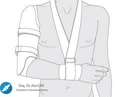
Treatment may be conservative or surgical, depending on the type of fracture, the age of the patient and the accompanying complications. In non/minimally displaced fractures: immobilization with shoulder-arm sling and passive ROM are applied. While the aim is to save the humeral head in young patients; Hemiarthroplasty option should be considered in elderly patients with osteoporotic and degenerative complaints.
Surgical treatment is preferred in cases where conservative treatment is insufficient, in cases of severely displaced humeral head or dislocation, in cases of plexus brachialis injury and in multi-part fractures of the humeral head. Complications: Neurovascular injuries, myositis ossificans, frozen shoulder, avascular necrosis, malunion and nonunion. Avascular necrosis is common in displaced 3-piece fractures, 4-piece fractures, and those with T plate application.
HUMERUS BODY FRACTURES
Humerus shaft fractures It occurs due to direct or indirect trauma. It usually breaks due to direct trauma. It fractures transversely and in pieces with direct trauma, and obliquely and spirally with indirect trauma. It is common in young adults. If the fracture is above the attachment point of the deltoid muscle (junction of 1/3 upper and 1/3 middle), the lower part is pulled laterally, and the upper part is pulled by the muscles coming from the chest and back.
slides inward. If the fracture site is below the attachment point of the deltoid muscle, the lower part is displaced upwards and the upper part is displaced outwards and forwards.
In his clinic, there is pain, swelling, ecchymosis and limitation of movement. Radial nerve paralysis is common in humerus fractures and 1/3 mid-distal region fractures, but since the majority of cases are neuropraxia, spontaneous recovery occurs. Fractures of the middle-lower 1/3 junction of the humeral body are generally spiral and the fracture line goes from lateral superior to medial-inferior. this type humerus fractures During reduction, the radial nerve may become trapped between the fragments.
Conservative or surgical treatment is applied in the treatment of humerus fractures. Conservative treatment may be preferred for non-displaced, slightly displaced or multi-fragmented fractures. Conservative treatment methods are divided into two.
1) Thoracobrachial Immobilization: Velpau bandage, shoulder strap, thoracobrachial spica cast.
2) Dependency Traction: These are methods that provide alignment control using gravity. Hanging arm cast, U splint, Sarmiento brace (Functional brace).
The purpose of the suspended plaster method is to apply traction to the fracture by using gravity to ensure alignment. The cast is applied with the elbow flexed at 90 degrees. The plaster cast must be positioned well on the arm. In the U splint: When the elbow is flexed at 90 degrees, a splint is made that starts from the axilla, crosses the elbow and covers the deltoid.
Sarmiento brace is the preferred conservative treatment method in most cases and in many centers.
 Surgical Treatment Indications:
- More than adequate degree of angulation)
- Patients with multiple trauma where early mobilization is required (spinal cord injury, multiple long bone fractures)
- segmental fractures
- Pathological fractures
- Fractures with major vascular injuries
- Distal nerves that may develop radial nerve paralysis after closed reduction and manipulation humerus fractures (Holstein-Lewis)
- Ipsilateral radius and ulna fractures (floating elbow)
- Bilateral humerus fracture
- Brachial plexus palsy
- Neurological diseases such as Parkinson's and epilepsy
HUMERUS LOWER END FRACTURES
It may occur due to direct or indirect trauma. Fractures can be classified according to anatomical localization, as well as intra-articular, extra-articular, intra-capsular and extra-capsular fractures. A careful neurovascular examination is required.
Treatment: For good results, anatomical joint and mechanical axis reduction and rigid internal fixation that allows early movement are provided. Internal fixation usually consists of two plates and screws placed posterior to the radial column and medial to the ulnar column. Primary total elbow prosthesis constitutes an alternative for comminuted fractures in elderly patients.
SUPRACONDYLAR HUMERUS FRACTURES
These fractures are generally summer fractures. The most common form occurs by falling on an open hand, with the elbows in hyperextension. The distal humerus is broken just above the physis. Since edema may develop in a short time, following the reduction, it is first splinted and monitored. There may be a risk of slipping and complications due to swelling; It may result in angular deformities or Volkmann's ischemic contracture. The lower part is pulled up and back with the influence of the triceps, and the upper part is pulled down and forward with the effect of the biceps. The upper part may damage the brachial artery and median nerve in the antecubital region.
supracondylar humerus fractures It is the most common elbow fracture in children. It has great clinical importance because it is the most common cause of Volkmann ischemic contracture. These fractures are divided into two groups: extension type (distal fragment in the posterior) as a result of falling while the elbow is in hyperextension, and flexion type (distal fragment in the anterior) as a result of falling while the elbow is flexed.
Ekstansiyon tipi kırık daha sık görülür (%95). Klinikte ağrı, şişlik ve hareket kısıtlılığı görülür. Tedavide kaymamış kırıklarda dirsek 90 derece fleksiyonda ön kol pronasyonda uzun kol alçısı yapılır. 4-6 hafta tesbit tedavi için anestezi altında kapalı yeterlidir. Hafif deplasman varsa redüksiyon ve dirsek 90 derece ve ön kol süpinasyonda alçı yapılır. Tam kaymış kırıklarda devamlı iskelet traksiyonu veya cerrahi uygulanır.


 Types of Humerus Fractures
 Surgical Treatment Indications:
Hello teacher. I need your help on a very important matter, so I wanted to contact you with your permission. Sir, I had surgery on my arm in 2009 and a postoperative diagnosis was made as "OPERATED LEFT HUMERUS MEDIAL CONDYLE FRACTURE". I am currently in the police school and there is an article in the police health regulation that says "Extra-articular plates and foreign objects placed for the detection of fractures that do not determine the description and do not disrupt joint movements and functions if removed are accepted as students." However, I do not know whether this operation is intra-articular or extra-articular. If it was intra-articular, I would be eliminated. Please help me sir, I will do whatever you want.
Finally, what was done during the surgery is written as follows in the report: "THE TRAILER WAS REACHED WITH AN INCISION OF APPROXIMATELY 3 CM OPEN ON THE MEDIAL CONDYLE, DETECTION WAS PROVIDED WITH 2 K WIRES, A CONTROL FILM WAS TAKEN AND ENDED WITH DRESSING.
SIR, CAN YOU PLEASE TAKE A LOOK AT IT, IS THIS AN OPERATION PERFORMED INTO THE JOINT?
Great post that gives insightful knowledge by giving the percentage details.The treatment process was well explained.